When the locum job was confirmed I was scared stiff. Being nervous when going into a new placement is common but this is not common practice. The practice is a cardiac referral practice, the best in Europe, ok let’s not beat about the bush, the best in the world.
This practice does not follow some ones else’s animal cardiac textbook, they wrote it, literally. Small animal cardio/ respiratory work done elsewhere in the world was pioneered and perfected here. They still write major scientific papers that change the course of thinking and treatment of small animal cardiology, world-wide.
As it turns out l need not have worried. I felt at ease from the start. One thing that did it was the relaxed atmosphere, the other is that much of the work is standard veterinary nursing work.
Looking after inpatients, reception, phone calls, anaesthesia, x rays (although machines do vary they are fairly easy to get to grips with) topping up, checking what to order, general cleaning.
With equipment l haven’t used, or procedures l haven’t done, or not sure on everyone is great at explaining how to do/use whatever. I just wish l could learn faster, l guess it is me getting old.
A lot of ultrasound scanning is done. Before now an ultrasound tended to be a shifting mass of grainy images to me. Now lm getting good at spotting things. I can recognise a heart valve when l look at it, don’t know which one it is in the heart, but hey, it is a start.
Both vets are great at explaining what is showing on the screen, how it works/isn’t working and why it may be like that (so often genetic), and what may need to be done, if anything is possible to correct the problem.
We had one very sweet little bitch having her scan done and she was hooked up with 3clips to her legs to check her basic ECG. Initially her tail kept doing gentle intermittent little wags, but she was totally still otherwise.
“Ahhh said the vet, l think l know what’s going on” He undid the clips and taped them to her feet.
The waggy tail stopped and she did not move till we turned her round to scan the other side of her heart, when she immediately lay down and totally still.
The clips had been a bit to much for her but she was such a sweet little lady she did not wriggle, howl, cry or use any other rude way to explain the problem. Just a gentle intermittent wag of the tail, Her polite way to say “excuse me please they hurt”
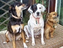
Unlike the young nutty hooligan dog who did not want to lie down when there was so many exciting things going on.
He felt that it was the height of insult to have jelly stuff on him that he was not allowed to eat. He only had the humans word for it that ultra sound gel is not tasty he wanted proof.
Then there was all the new stuff that he wanted to sniff, and given the chance pee on, to prove he had been there!
I have what looks like another fine set scars forming from his nails on my arm to remind me of him.
When animals are like that then they are sedated lightly to settle them. Mind you that behaviour and effect is normal in all practices not nasty just overexcited.
It is safe to say that several operations here have “blown my little mind” as the well known saying goes. I am used to major operations in animals but these are major delicate operations. Using equipment that you just do not see in general or even general referral practice.
The effects so far are also very different. Instead of often intense nursing and several days recovery these patients scoffed a good meal that night and were home the next day.
Operations here are often done guided by fluoroscopy. These are x-ray pictures that allow you to see internal organs or procedures live and in real time. The fluoroscopy has quite a few uses.
We used it on a dog that was drinking liquid barium and eating barium mixed with dog food. This was to observe the swallowing action to try and determine where the problem was, and what caused/contributed to the patients cough.
The other diagnostic aids had not been very clear and were of limited use in diagnosing this patients problem.
Amazing to see what you have learnt happens via a textbook about the body’s mechanisms when something is swallowed, actually happening live.
I know l have seen it in documentaries but that is remote, this was not.
Operations wise, for the following 2 especially, there are both of us veterinary nurses. I do what l know for anaesthetics and leave the complex assist parts and equipment to the head nurse. With her in throat clearing distance if l needed anything.
People could learn a hell of a lot from her, she is calm, cheerful, knowledgeable and helpful. In this profession more are needed like that behind the scenes.
Fluoroscopy was used in 2 different types of heart operations this time the patients were under general anaesthetic.
PDA
The first, a large dog about 35kgs in weight, had a PDA or “hole in the heart” repaired.
**Patent ductus arteriosus (PDA) is a when a vessel connecting the two major cardiac vessels (the aorta and the pulmonary artery) fails to close at birth as it should. Uncorrected, a PDA leads to progressive heart enlargement and heart failure. If left untreated, approximately 50% of dogs with a PDA die in the first year of life.
The closure involved passing catheters via the artery in the hind leg (‘keyhole’ surgery) and ‘plugging’ the vessel with devices to stop or minimise flow through it. This was also assisted with angiographic studies*
Visually post operation there was a small wound where the incision and that was it. There is minimal pain compared to traditional surgery (which involves cutting open the chest between the ribs).
That evening l was walking the dog and trying to stop her bouncing about and jumping up for kisses with me, not easy she could see no reason to keep calm. She went home the next day.
**In some cases where traditional “open chest” is needed here, patients are hospitalised for 3 - 5 days to provide pain relief and to monitor for complications that can arise following open chest surgery. After discharge dogs must be rested for 3 or 4 weeks so as not to pull any of the sutures, especially those between the ribs. After that, exercise can be gradually introduced over a period of a couple of weeks.
A huge contrast to the method l witnessed.
BV
The second operation was a Balloon Valvuloplasty on a small cross breed about 10kgs in weight.
She had Pulmonic Stenosis. **This congenital heart condition restricts blood flow through to the pulmonary artery, which takes blood to the lungs for oxygenation. As a consequence of this narrowing, blood flows through the valve at an increased velocity. This results in a ‘squirt’ of blood flow with each heart beat, which creates a sound called a murmur. The obstruction caused by the narrowed valve leads to an increased pressure in the right heart, and potentially heart failure
The operation to repair it “Balloon valvuloplasty” involved passing a specially designed balloon-tipped catheter via the vein in the neck (‘keyhole’ surgery) into the heart and through the narrowed valve.
This is catheter is visually guided by fluoroscopy and then after *angiographic studies. A sausage-shaped balloon was momentarily inflated to stretch the defective valve, allowing it to open more normally.
Again post op all she had to show for this major operation was a small wound on her neck and a clipped area of her coat.
That night the she out for a walk. She was not to happy, it was dark, windy and wet, who want’s to walk in that? Again she went home the day after her operation.
They also carry out pacemaker operations here as well, although l have not seen any, yet.
*====*====*====*====*====*====*====*====*====*====*====*====*
*An angiogram is a radiographic technique used to visualise blood vessels (the fluoroscopy can not show these). A dye is injected into the heart arteries with a special catheter. This dye enables the fluoroscopy to contrast the arteries with the surrounding body tissue. The cardiologist can then see the artery even the smallest blood vessels. (The angiogram was another first for me)
**Explanations of PDA and Pulmonic Stenosis were 'borrowed' from the practice website. My words are mixed in with them.
Subscribe to:
Post Comments (Atom)


1 comment:
Mike seemed quite chuffed and told me that he has posted "l has been blogged" on the vetsurgeon site.
He has not, as far as l know read the blog although l did clear permission with him first.
Post a Comment